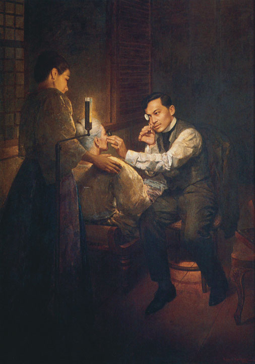
 Today, June 19 is the 159th birthday of our National Hero Jose Rizal. This famous painting above shows Jose Rizal, who trained as an ophthalmologist in Europe in the 1880’s, examining his mother. He is doing a procedure that is still commonly done by all ophthalmologists to this day, albeit with more sophisticated instruments. He is looking at her retina and optic nerve, The retina is an important part of at the eye. It is the innermost layer and it contains the cells that are able to detect light and transform it into nerve impulses which in turn is sent to the brain, allowing us to see. Being such an essential part of the eye, it is very important that the retina be examined closely. But, it’s not that easy. Since the retina is located inside the eye and the only opening, the pupil, is less than half a centimeter, the inside of the eye is normally in near or total darkness. To see the retina, the examiner must provide a source light as illumination otherwise it will be too dark to see anything inside. The tiny pupil limits how much light can enter the eye at any given time to protect the light-sensitive retina from being overwhelmed. This is why you instinctively blink or even turn away to avoid the glare when someone flashes a very bight light in front of you and why it is dangerous to look at the sun directly. To make things even harder, the examiner has to peer through the same pupil as that of the source of light, so his line of sight will necessarily have to be in the same path. In other words his line of sight as well as the light source have to be on the same axis, or are coaxial. (see fig.1) 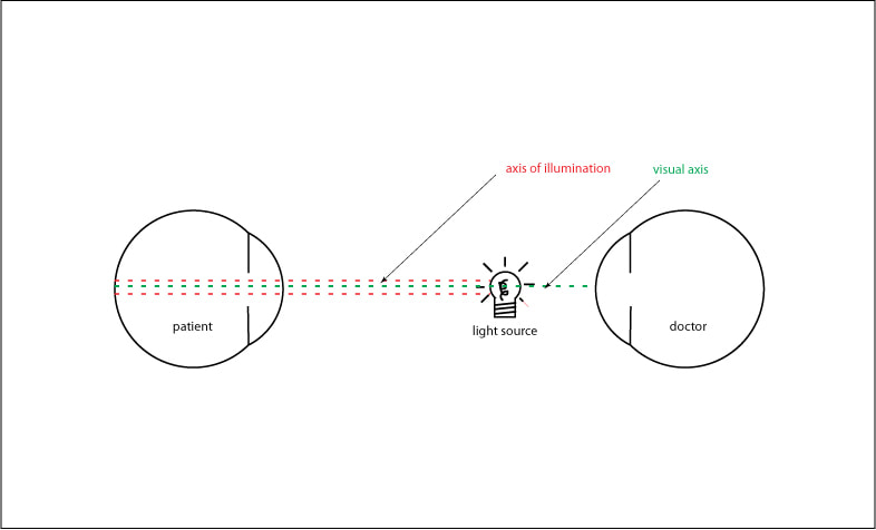 fig 1: the line of sight and the source of illumination have to be on the same axis (co-axial) in order to see the retina In the painting, the source of light is behind his mothers head. Rizal is holding with his right hand a small concave mirror with an opening in the center. This reflects and focuses the light into a narrow beam that is then directed to the inside of the eye through the pupil. The image of the retina is visible on the condensing lens that he holds in his left hand. Since he is looking through the opening in the center of the concave mirror, his line of sight is now co-axial with his examining light. 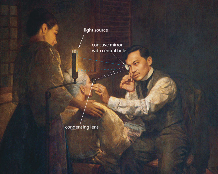 Nowadays ophthalmologists, and especially retina specialists still use similar instruments that rely on the same principle albeit more sophisticated thus making the procedure much easier to perform and with less discomfort for the patient and the examiner. Examining the retina is a very important in a lot of diseases because it allows the eye specialist to see in real life the actual condition of the blood vessels and nerves inside our bodies which otherwise would not be visible. Retina specialists are highly skilled in peering deep inside the eye and enables them to see such conditions as retinopathy of prematurity, ARMD, diabetic retinopathy, retinal detachment, vitreous hemorrhage and many more. Glaucoma specialists and neuro-ophthalmologists also rely on this to see status of the optic nerve. In a way, it allows us a peek into the human brain. 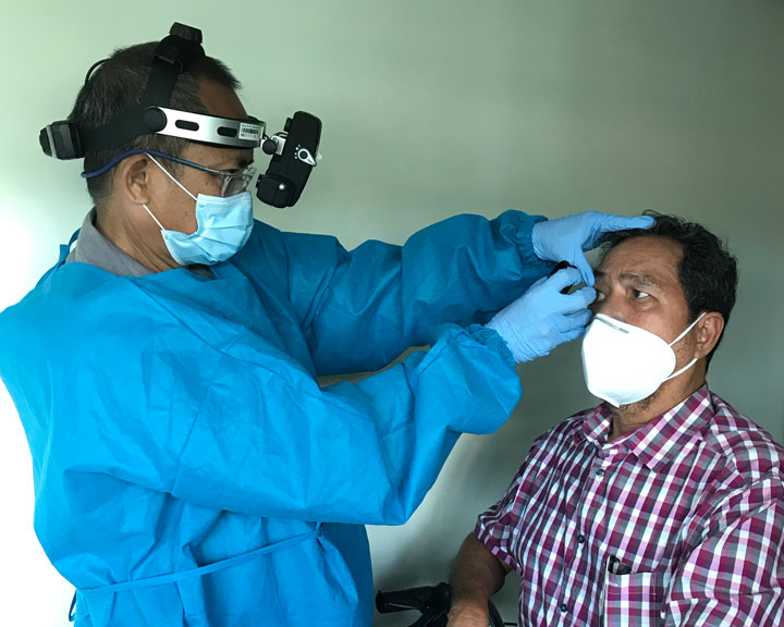 Comments are closed.
|
Aces CaresOur Stories Archives
February 2021
Categories |
 RSS Feed
WORLD-CLASS EYE CARE IN THE HEART OF THE PHILIPPINES
|
What Our Clients Are Saying"experience ko very affordable! you are not checked by one doctor only, but by every specialist they have!!, i had cataract at age 52, the examining doctor suspects i may develop glaucoma so he had me checked by their glaucoma specialist. when i complained of dryness in the eyes, another specialist examined me. btw, i was initially examined and operated on by a retina specialist of theirs all went well!!"
Brought my mom there for removal of cataract... Our experience with this clinic and with Dr Aquino who was mom's surgeon was nevertheless outstanding..." "I was here years back...The doctors who attended to me were very accomodating and were even able to build well a doctor-patient relationship..."

photo: Arambulo
|